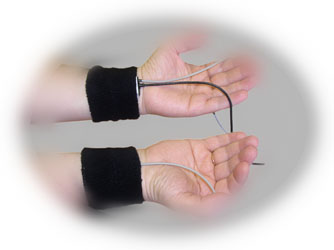
| Photoplethysmograph (PPG) was developed in the 1960's and 1970's by psychophysiology researchers. The instrument is used for assessment of peripheral blood flow to the extremities. When the client is stressed their blood vessels constrict (less blood flow in fingers). Photoplethysmograph is based upon the premise that all living tissue and blood have different light-absorbing properties. PPG works by placing an individual finger between two parts of a transducer consisting of a light source and a photocell (which converts light to electrical energy). A beam of infrared light is projected toward the photocell. The blood in the finger scatters light in the infrared range, and the amount of light reaching the cell is inversely related to the amount of blood in the finger. Hence, when blood vessels in the finger dilate, the increased blood flow allows less light to reach the photocell, when blood vessels constrict, blood flow is decreased and increased light reaches the photocell. Pulse volume measurements are related beat-to-beat variations in the force of blood flow. Phasic changes made on a beat-to-beat basis, are called pulse volume measurements and are related to beat variations in the force of blood flow. These beat-to-beat changes in peripheral blood flow reflect ANS regulatory activity and can be used for the purpose of evaluation of sympathetic activity or stress. (Measuring Stress by S. Cohen, R. Kessler and L. U. Gordon, 1995) |